


*****The following content was written by the author(s) of the paper.*****
A joint research group consisting of Hikaru Ichida, a doctoral student in the Division of Nano Life Science, Graduate School of Frontier Science Initiative, Kanazawa University; Kosuke Mizuno, currently a Postdoctoral Researcher at the Institute for Protein Research, The University of Osaka; Professor Noriyuki Kodera and Associate Professor Holger Flechsig of the Nano Life Science Institute (WPI-NanoLSI), Kanazawa University; and Associate Professor Satoshi Toda of the Institute for Protein Research, The University of Osaka, has succeeded in visualizing the structural dynamics underlying how the serum protein Afamin stabilizes and transports Wnt3a, a lipid-modified signaling molecule. The study also showed that stable binding between these two molecules depends on both a hydrophobic pocket (*1) that accommodates Wnt3a and the structural integrity of Afamin.
Wnt proteins are essential molecules that help the body develop properly and maintain healthy tissues. However, because they do not dissolve well in water and are highly hydrophobic, they tend to be unstable in the body. This study has revealed part of the mechanism by which Wnt3a is stably transported with the help of another protein.
These findings are expected to deepen our understanding of biological processes involving Wnt3a and may contribute in the future to the development of ex vivo tissue engineering technologies and regenerative medicine.
The results of this study was published in the online edition of "Nano Letters" on April 15th, 2026.
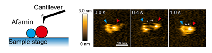
Fig.1: Schematic illustration of high-speed AFM (*2) observation (left) and consecutive high-speed AFM images of Afamin (right). Afamin consists of a large part (red) and a small part (light blue), and was observed to flexibly expand and contract.
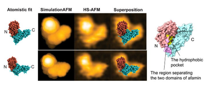
Fig.2: Analysis of high-speed AFM images using a computational method to estimate the protein’s three-dimensional atomic structure model (left), and the Afamin structure color-coded based on the analysis results (right). The region shown in pink is an unstructured region, and the study revealed that the center of this region can be used to distinguish the N-terminal domain from the C-terminal domain. The hydrophobic pocket, shown in yellow, is located near the center of Afamin.
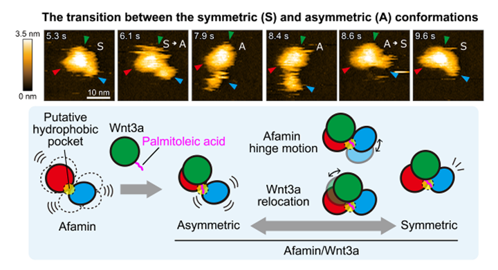
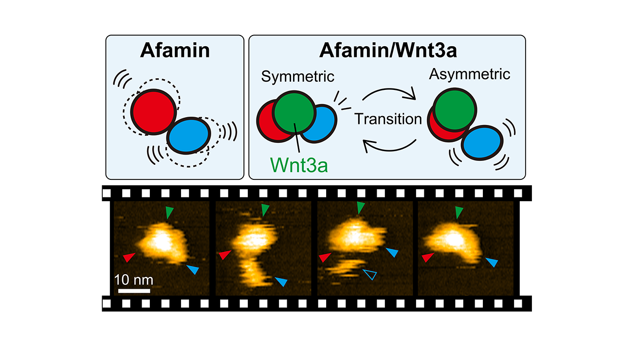
Fig.3: Consecutive high-speed AFM images of Afamin/Wnt3a (top) and a model of its motion (bottom).
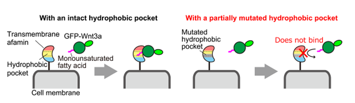
Fig.4: Schematic illustration of the binding experiment using Afamin mutants fixed on the cell surface. Wnt3a binds to Afamin with an intact hydrophobic pocket (left), but does not bind when part of the pocket is mutated (right).
【Glossary】
*1 Hydrophobic pocket
A structural region that can accommodate molecules that do not dissolve well in water. In this study, this pocket was found inside Afamin and shown to play an important role in holding the lipid part of Wnt3a.
*2 High-speed AFM
A unique microscopy technique that can capture the shapes and movements of biomolecules such as proteins in liquid as video. It can record molecular motion in real time with a time resolution of about 0.1 seconds (10 frames per second). Its spatial resolution is about 1 nm in the horizontal (XY) direction and about 0.1 nm in the vertical (Z) direction, which is high enough to distinguish protein domain structures.
Click here to see the press release【Japanese only】
Journal:Nano Letters
Researcher Information : Noriyuki Kodera
Holger Flechsig
Related Information
Graduate School of Frontier Science Initiative, Kanazawa University
Nano Life Science Institute, Kanazawa University