


金沢大学医薬保健研究域保健学系 市川勝弘教授は、名古屋市立大学医学部附属みどり市民病院(名市大みどり市民病院)放射線技術科 大橋一也技師⾧との共同研究により、内視鏡(胃カメラ)を使わずに胃の内部を鮮明かつ立体的に可視化する「バーチャル内視鏡検査法」を開発しました。本検査法は、2 管球 CT 装置で得た CT 画像に独自開発の画像処理を組み合せ、まるで胃の中で目視しているかのような没入的画像観察手法です。これにより次世代の胃がん検診の確立を目指します。従来のバリウム検査に伴う誤嚥や便秘のリスクを排除し、内視鏡専門医不足という社会的課題を解決する、体への負担が少ない新しい検診スタイルの提案です。
本成果は 、2026 年 3 月 20 日の第 8 回日本消化管バーチャルリアリティ学会にて発表しました。
【ポイント】
- 「飲む」から「撮る」へ:バリウムやカメラの挿入が不要な、苦痛の少ない胃がん検診を実現します。
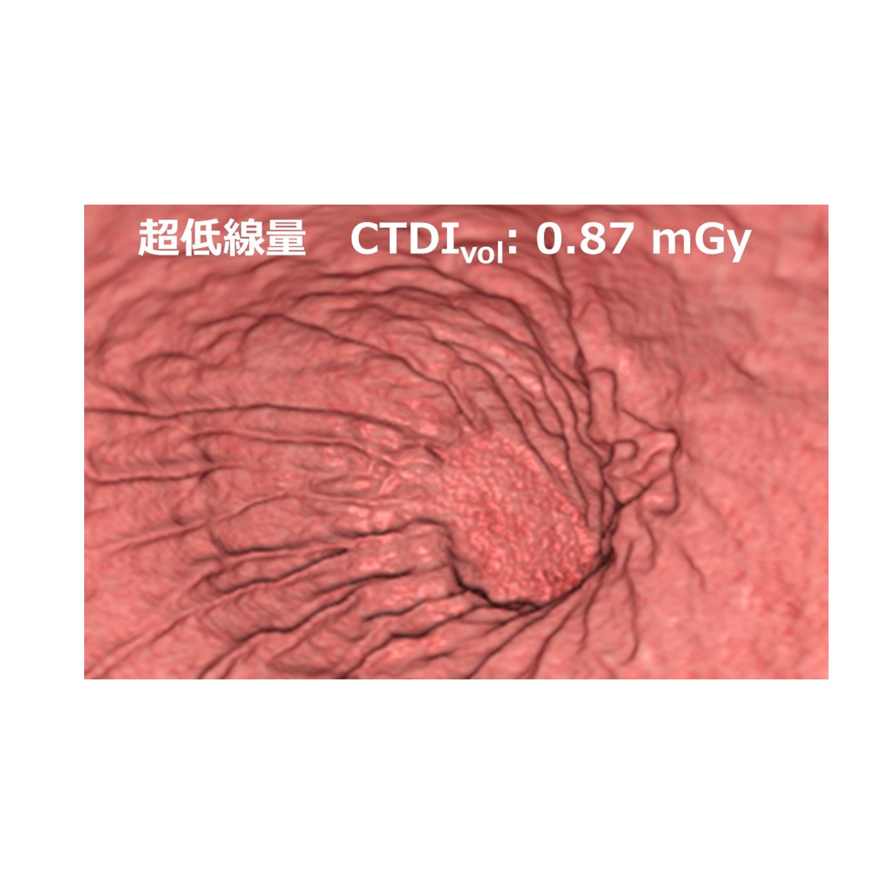
- 被ばく線量を大幅低減:プレフィルタ技術(※1)により、従来の胃 X 線検査(バリウム)よりも低い放射線量での検査を追求します。
- 死角のない 3D 画像:独自開発の写実的レンダリング技術により、実物に近い質感の3D 画像で胃壁をくまなく診断します。さらに3次元データにより胃カメラでは不可能な粘膜下の観察も可能です。
- 一回の検査で腹部のチェック:胃の表面だけでなく、肝臓・膵臓など上腹部の臓器も同時に検査可能です。
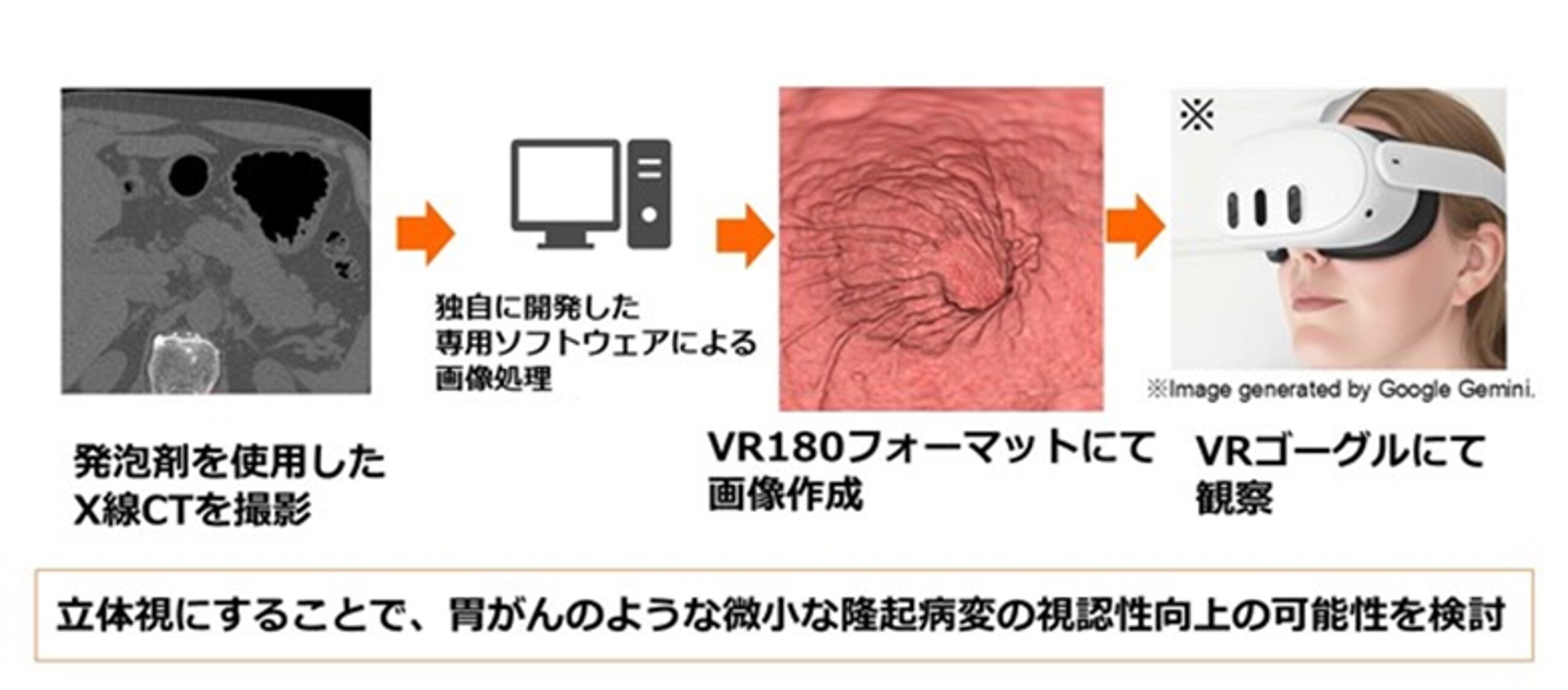
図1. 写実的仮想胃内視鏡(Photo-realistic virtual gastroscopy: PRVG)によるバーチャルリアリティ(Virtual reality: VR)胃検査システムの概要
【用語解説】
※1 プレフィルタ技術
胃と空気のようなコントラストが高い組織に対して、不要な低いエネルギーの X 線をカットすることで被ばく低減が可能な技術です。
研究者情報:市川 勝弘
関連情報
金沢大学 医薬保健学域 保健学類 / 大学院医薬保健学総合研究科 保健学専攻