




*****The following content was written by the author(s) of the paper.*****
A research group led by Professor Katsuhiro Ichikawa (Faculty of Health Sciences, Institute of Medical, Pharmaceutical and Health Sciences, Kanazawa University) and Professor Kaoru Tada (Faculty of Health Sciences / Department of Orthopaedic Surgery, Kanazawa University Hospital) has successfully developed an ultra-high-resolution X-ray computed tomography (CT) system dedicated to extremities, achieving approximately five times higher spatial resolution than conventional X-ray CT systems (voxel size: 0.08–0.10 mm).
The spatial resolution of X-ray CT systems commonly used in clinical practice is approximately 0.5 mm, which is insufficient for visualizing the fine internal bone microstructures known as trabeculae, whose thickness is typically 0.1–0.2 mm. To overcome this limitation, the research group conducted extensive investigations into the scanning mechanisms of CT systems, including the X-ray source, detector, and rotational drive mechanisms, and optimized them to a level capable of clearly depicting trabecular bone in the extremities.
As a result, the voxel size—representing the three-dimensional unit of reconstructed data—was successfully reduced to 0.08 × 0.08 × 0.08 mm³. Previously, CT systems with such extremely small voxel sizes were limited to experimental setups that could not be applied to the human body. The newly developed system enables very short-time scanning of the extremities, imposes minimal burden on subjects, and nonetheless provides clear visualization of fine bone microstructures. The system has already been completed in a configuration suitable for clinical application, and it is expected to contribute to accurate diagnosis of bone diseases and traumatic injuries of the hands and feet in the future.
This research was published online on November 13, 2025, in the specialized journal Skeletal Radiology.
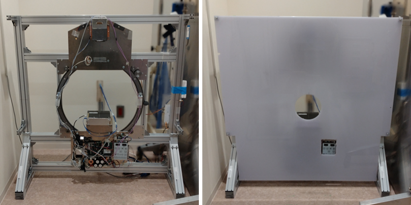
Figure1.Internal configuration (left) and external appearance with the front panel attached (right) of the ultra-high-resolution X-ray CT system.
The system consists of a compact X-ray source, a high-resolution X-ray detector composed of 0.099-mm detector elements, and a computer-controlled motor-driven gantry that rotates these components with high central alignment accuracy. CT scanning procedure is performed while rotating the gantry and emitting X-rays, and the acquired projection data are reconstructed into three-dimensional images with voxel sizes of 0.08–0.10 mm.
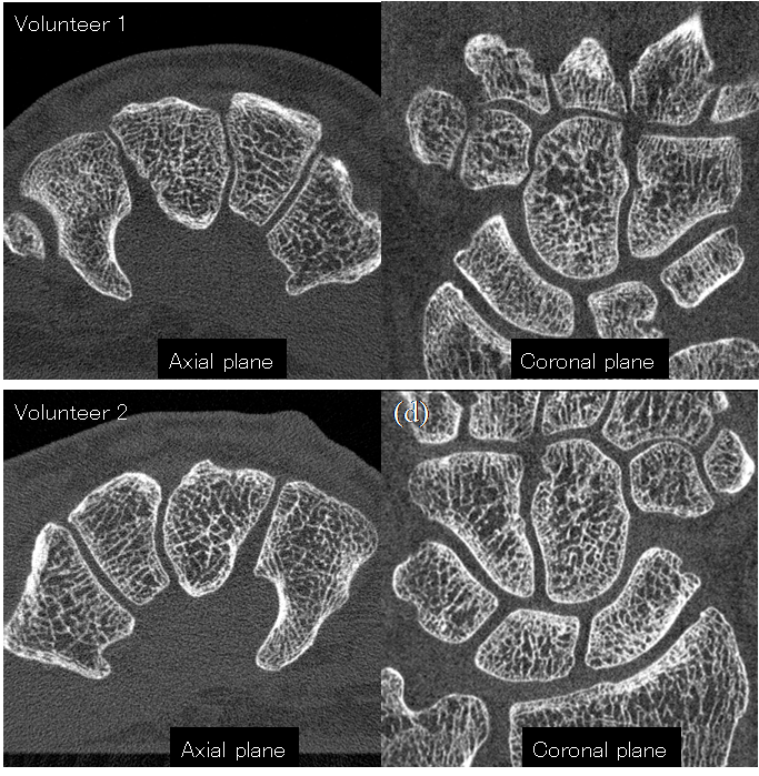
Figure2.CT images of the hands (carpal region) of two healthy volunteers acquired with the developed system.
The internal fine bone microstructures (trabeculae) of the carpal bones and wrist joints are clearly and almost completely separated and visualized. Together with the axial images, the coronal image reconstructed parallel to the palm confirms that ultra-high spatial resolution is maintained in all three dimensions.
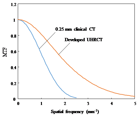
Figure3. Comparison of spatial resolution characteristics between the developed system and a clinically available CT system equipped with a 0.25-mm detector.
In the graph, curves extending further to the right and higher upward indicate superior spatial resolution. The developed ultra-high-resolution CT system clearly outperforms the CT system using a 0.25-mm detector.
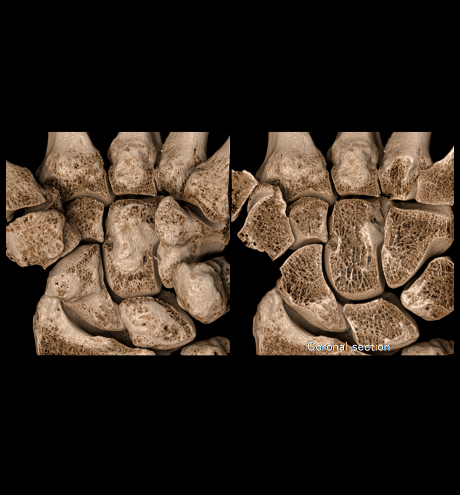
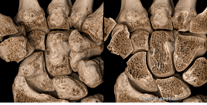 Figure4. Highly realistic three-dimensional image generated using a novel visualization technique developed in parallel with the CT system.3
The technique enables the generation of images that appear as if a physical specimen were extracted from the living body. Although ultra-high-resolution CT data consist of a very large number of slices, this visualization method allows comprehensive observation at a glance and enables virtual sectioning to inspect internal structures as needed.
【Glossary】
*1 X-ray Computed Tomography (CT) System
A medical imaging system that uses X-rays and computer-based calculations to generate images of the internal structures of the human body. In conventional X-ray radiography, internal organs and tissues overlap in a single projection image and cannot be clearly separated. In contrast, a CT system reconstructs cross-sectional images, allowing internal structures to be visualized separately and in detail.
*2 Spatial Resolution
A measure of how small a structure can be distinguished as separate objects in an image. For example, a spatial resolution of 0.1 mm means that multiple structures of approximately 0.1 mm in size can be individually recognized even when they are located close to each other.
*3 Trabeculae
Fine, interconnected pillar- or beam-like structures within bone that form a complex internal network. Similar to the beams of a building, trabeculae support the bone from the inside and play a critical role in maintaining bone strength.
*4 CT Scanning
The process of acquiring imaging data by irradiating the human body with X-rays while recording the intensity of X-rays transmitted through the body. These data are then used to reconstruct cross-sectional and three-dimensional images.
Click here to see the press release【Japanese only】
Journal : Skeletal Radiology
Researcher Information : Katsuhiro Ichikawa
Kaoru Tada
Related Information
 PAGE TOP