


Abstract:
Aire-expression in the thymus is crucial in immune tolerance by eliminating auto-reactive T cells; insufficient elimination causes autoimmune disorders. Aire was thought to be expressed only in the thymus but recently Aire-expression in peripheral lymph nodes was revealed. We identify the cells expressing Aire in lymph nodes, 0.01% of lymph node cells, to be ILC3-like cells using Aire-reporter mice and flow cytometry. The results may lead to elucidation of causes of autoimmune disorders and treatments.
[Background]
The thymus is the organ in which T cells*1) differentiate. T cells express various T cell receptors for reacting with various pathogenic microbes and viruses. However, the thymus generates auto-reactive T cells that react with self antigens, which must therefore be eliminated in the thymus. The mechanism for elimination of auto-reactive T cells is referred to as central tolerance*2). Medullary thymic epithelial cells play key roles in central tolerance. These cells express organ-specific antigens (tissue-restricted antigens). For example, insulin*3) is produced only by pancreatic β cells and C-reactive protein*4) is produced only in the liver. The medullary thymic epithelial cells produce and present such proteins to T cells and eliminate those T cells that react with them, i.e. auto-reactive T cells (Figure 1). Aire*5) is a gene that regulates expression of tissue-restricted antigens in medullary thymic epithelial cells. Mutation of Aire causes decreased expression levels of tissue-restricted antigens in the thymus, and consequently, elimination of auto-reactive T cells becomes insufficient. This is thought to lead to autoimmune disease*6) pathogenesis in multiple organs such as ACEPED*7). It was believed that Aire was expressed only in the thymus, but several research groups recently presented evidence that some cells in peripheral lymph nodes express Aire; peripheral lymph nodes are the organ where immune reactions take place. However, there is disagreement between these research groups on Aire-expressing cell types.
[Outline of research results]
A scientist from Kanazawa University played central roles in this immunological study as a member of an international collaborative research group, which has been engaged in studies on the mechanism of central tolerance induction in the thymus. During the course of their research, the group found Aire gene expression in lymph nodes; Aire gene had been thought to be expressed only in the thymus. Thus, cells expressing the Aire gene in lymph nodes were analyzed in detail using two strains of Aire-reporter mice*8) and high sensitivity flow cytometry*9) using Aire antibody. This confirmed that a very small number of cells, 0.01% population of all the cells in lymph nodes, did indeed express Aire. These cells were found to express a high level of major histocompatibility complex (MHC)*10) class II, which is expressed on antigen presenting cells. They also showed a high level of expression of costimulatory molecules necessary for T cells to be activated, which suggested Aire-expressing cells might play roles as antigen presenting cells. These Aire-expressing cells were analyzed in terms of cell morphology, cell surface molecules and gene expression profile, by which these cells were found to show characteristics similar to those of group 3 innate lymphoid cells (ILC3)*11) that play important roles in maintaining homeostasis and the inflammation reaction of the gut. The present research group named these cells Aire ILC3-like cells. A previous study detected Aire mRNA in a cell fraction, which was confirmed in this study, but the cells in the fraction expressing Aire protein were Aire ILC3-like cells only.
Next, the gene expression profile of Aire ILC3-like cells was studied using wild-type mice and Aire knockout mice, which indicated that about 700 genes were regulated by Aire. In experiments using mice whose Aire-expressing cells were made to express hemagglutinin-antigen (HA antigen) of influenza virus, Aire ILC3-like cells were found to eliminate HA-reactive T cells in lymph nodes (Figure 3). These results indicate that Aire ILC3-like cells play a part of the role of immune tolerance in lymph nodes.
[Future prospects]
Immune tolerance failure leads to autoimmune diseases. It is considered that peripheral T cell tolerance in lymph nodes consists of suppression by regulatory T cells, elimination of auto-reactive T cells and induction of anergy*12) but it was unknown as to which types of cells were involved and in what manner in elimination of auto-reactive T cells and induction of anergy. Aire ILC3-like cells discovered in this study are thought to be involved in the elimination of a proportion of auto-reactive T cells. In the near future, we hope to elucidate how immune tolerance is maintained by Aire ILC3-like cells and other cells inducing immune tolerance. This should lead to elucidation of the causes and possible treatments of connective tissue disease and autoimmune diseases.
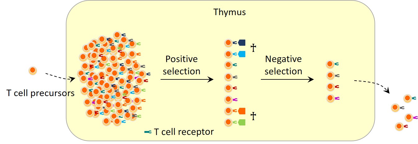
Figure 1. Auto-reactive T cells that attack self-antigens are eliminated in the thymus.
T cell precursors enter the thymus and generate a large number of TCR repertoire. Among TCR repertoire, there are some auto-reactive T cells. Auto-reactive T cells are negatively selected in the thymus, while self-tolerant T cells, after maturation completion, move to peripheral lymphoid organs such as lymph nodes and the spleen.
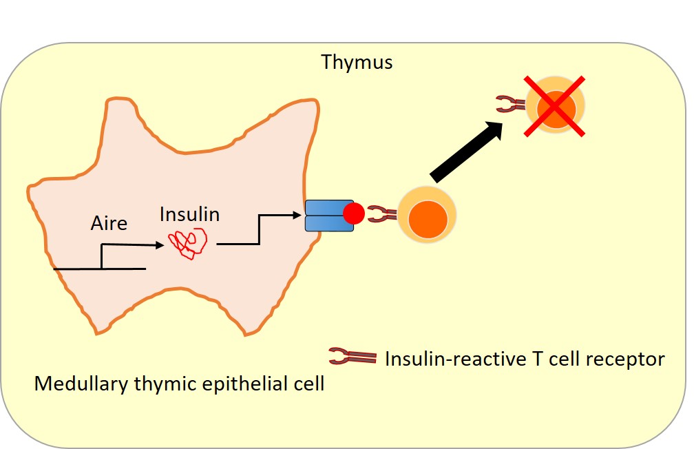
Figure 2. Aire induces the expression of tissue-restricted antigens in medullary thymic epithelial cells, contributing to elimination of auto-reactive T cells.
Medullary thymic epithelial cells express antigens that express in specific organs such as insulin, C-reactive protein, etc. Thus, it is possible to eliminate auto-reactive T cells, for example, those reactive with insulin.
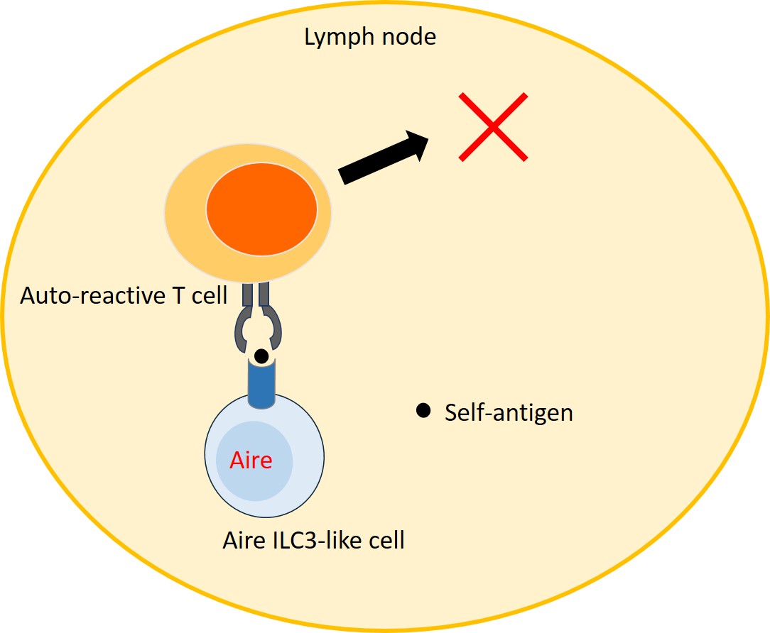
Figure 3. Aire ILC3-like cells eliminate auto-reactive T cells in lymph nodes.
It is shown in this study that Aire ILC3-like cells eliminate auto-reactive T cells in lymph nodes by presenting self-antigens.
[Glossary]
*1) T cell
A type of lymphocyte. T cell precursors enter the thymus where immature T cells differentiate into mature T cells, which then move to peripheral lymphoid organs such as lymph nodes and the spleen. T cells play essential and central roles in the immune system in eliminating cancer cells or cells infected by virus.
*2) Immune tolerance
Immune tolerance is a state of unresponsiveness of the immune system to specific antigens. Failures in immune tolerance cause autoimmune diseases. T cell immune tolerance is classified into central tolerance induced in the thymus and peripheral tolerance induced in peripheral lymphoid organs such as lymph nodes and the spleen.
*3) Insulin
A protein hormone secreted by pancreatic β cells. Insulin is secreted in response to increases in glucose levels and serves to lower them. Deficits of insulin induce a rise in blood glucose levels, causing the pathogenesis of diabetes. Type 1 diabetes is caused by auto-reactive T cells attacking pancreatic β cells.
*4) C-reactive protein
C-reactive protein is found in blood plasma in case of inflammation and tissue destruction.
*5) Aire
Aire (autoimmune regulator) is an essential gene for preventing autoimmune diseases. Aire is expressed primarily in medullary thymic epithelial cells, eliminating auto-reactive T cells by inducing expression of tissue-restricted antigens.
*6) Autoimmune disease
Autoimmune disease is a pathological condition arising from an abnormal immune response to normal body constituents. The immune system that works for protection from bacteria and viruses attacks the self antigens, which leads to autoimmune diseases. Autoimmune diseases include rheumatoid arthritis, type 1 diabetes, multiple sclerosis etc.
*7) APECED
It is a genetic disorder inherited in autosomal recessive fashion due to a mutation in the Aire gene, whose major symptoms are organ-specific autoimmune disease and mucocutaneous candidiasis.
*8) Aire-reporter mouse
Genetically manipulated mouse that expresses fluorescent protein in the Aire-expressing cells.
*9) Flow cytometry
Flow cytometry is a technique used to detect and measure physical and chemical characteristics of a population of cells such as cell size and surface antigens. A suspension of cells, ideally in a single row, flows into the flow cytometer, where each cell is irradiated with a laser beam. The light scattered by each cell is analyzed to measure the abovementioned properties.
*10) Major histocompatibility complex
The major histocompatibility complex (MHC) is a set of genes that code for cell surface proteins essential for the acquired immunity in vertebrates. MHC class II molecules are found only on antigen-presenting cells such as dendritic cells, thymic epithelial cells, and B cells. These cells are important in initiating immune responses.
*11) Innate lymphoid cells (ILCs)
Innate lymphoid cells (ILCs) were recently identified as a new group of lymphocytes, defined by the absence of antigen receptors. They consist of group 1 ILCs (ILC1), group 2 ILCs (ILC2) and group 3 ILCs (ILC3), regulating the immune system by secreting various cytokines. ILC3 is primarily found in the gut lamina propria, functioning in the maintenance of the epithelial cells and sustaining homeostasis with gut microbiota.
*12) Anergy
Anergy is an immune tolerance mechanism induced by the peripheral immune system. In anergy, T cells do not react with specific antigens.
Article
Aire expressing ILC3-like cells in the lymph node display potent APC features
Journal: Journal of Experimental Medicine
Authors: Tomoyoshi YAMANO, Jan DOBEŠ, Matouš VOBOŘIL, Madlen STEINERT, Tomas BRABEC, Natalia ZIETARA, Martina DOBESOVA, Caspar OHNMACHT, Martti LAAN, Pärt PETERSON, Vladimir BENES, Radislav SEDLACEK, Rikinari HANAYAMA, Michael KOLAR, Ludger KLEIN, and Dominik FILIPP
DOI: 10.1084/jem.20181430
Funder
JSPS Leading Initiative for Excellent Young Researchers.