


Abstract:
Using our in utero electroporation technique for ferrets, we investigated the axonal fibers in the developing cerebral cortex, where ferrets have two fiber layers; the inner axonal fiber layer projects contralaterally and subcortically, whereas the outer fiber layer sends axons to neighboring cortical areas. Furthermore, mice and ferrets were found to have unexpected similarities. Our results shed light on the cellular origins, projection patterns, developmental processes, and evolution of fiber layers in mammalian brains.
[Background]
The cerebrum plays the most important roles in the higher functions of the brain. In particular, the cerebral cortex*1), among other parts of the cerebrum, is essential. Humans have by far the most developed cerebral cortex among animals and it is thought that we have acquired specific abilities thanks to this. In addition, the cerebral cortex has received special attention, since various parts are involved in various brain diseases, psychiatric disorders and others.
The developing cerebral cortex of higher animals like humans contains two axonal fiber layers that transmit neural information and are, therefore, considered to be important in brain functions. The cerebral cortex of the mouse, the most commonly used model animal in research, was not found to have equivalents of the axonal fiber layers, which made mouse research on this subject very difficult. Thus, research on these fiber layers has been much retarded.
The present research group at Kanazawa University has been promoting studies using the ferret*2), since it is important to conduct research using higher animals with a more developed cerebrum, closer to that of the human than the mouse. Research techniques for the ferret were previously not available, so in 2012 and 2013 the group developed an appropriate technique, in utero electroporation, for use in ferrets at the gene level. They have thus led research into the brains of higher mammals including the development of disease model ferrets in 2015 and 2017.
[Results]
In the present study, the Kanazawa University group has mapped the fiber layers of the developing cerebrum of a higher mammal, the ferret, using its own unique research technique. They have also found an important clue to the evolution of these fiber layers. More specifically, the following three points have been established:
1. The two axonal fiber layers found in the human and monkey brain also exist in the ferret brain.
By introducing GFP (green fluorescent protein) into neurons in the ferret cerebral cortex, it was found that axons in two fiber layers are derived from the neurons of the cerebral cortex (Figure 1 upper right panel, indicated with [ symbols).
2. The two axonal fiber layers have different destinations in the brain.
Upon investigation of the destinations of the two fiber layers, the one on the surface side of the cerebral cortex has destinations in the proximal areas of the cerebral cortex; i.e. it represents a short-distance pathway (Figure 1 lower panel, indicated with an arrow); on the other hand, the other in the deep side of the cerebral cortex has destinations in the cerebral cortex of the opposite hemisphere and to the other brain regions; i.e. it represents a long-distance pathway (Figure 1 lower panel, indicated by arrowheads). Thus, selection of the axonal fiber layers takes place depending on their destination.
3. A trace of axonal fiber bundles is found in the mouse brain.
So far, equivalents of the two fiber layers were not described in the mouse cerebral cortex. The group applied the same technique, used to reveal the two fiber layers in the ferret brain, to the mouse brain. Unexpectedly, they found one fiber layer (Figure 2, indicated by an arrowhead) as well as a trace of axonal fiber bundles (Figure 2, indicated by an arrow). This trace pathway is thought to have later evolved to become the second axonal fiber layer of higher mammals (Figure 3). This raises the possibility that it is this second axonal fiber layer, i.e. the outer fiber layer, which is important in the development of higher brain functions.
[Significance and future prospects]
In this study, the Kanazawa University group has elucidated the destinations of the two axonal fiber layers in the cerebrum and the process of their evolution with the use of their unique research technique for the ferret. This finding is of major significance, since there have been very few studies on these two fiber layers. This study should contribute to our understanding of brain evolution in higher organisms up to the human, which has been very difficult with the mouse, a conventional model animal. Further, it should help reveal causes of various brain disorders.
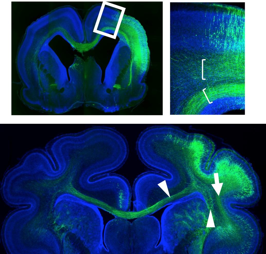
Figure 1. Two axonal fiber layers found in the ferret cerebrum and difference in their destinations.
Upper left panel. A cross-sectional image of the ferret brain. Some neurons of the cerebral cortex (blue) are dyed green with GFP, green fluorescent protein.
Upper right panel. The rectangle with white edges of the upper left panel is enlarged. It is found that there are two axonal fiber layers (green bundles) in the cerebral cortices.
Lower panel. The fiber layer on the surface side (green, indicated by an arrow), i.e., the outer fiber layer, has destinations in the proximal areas of the same cerebral cortex, while the deep fiber layer (green, indicated by arrowheads), i.e. the inner fiber layer, has destinations in the opposite cerebral cortex and other brain regions. Thus, the two axonal fiber layers have different destinations.
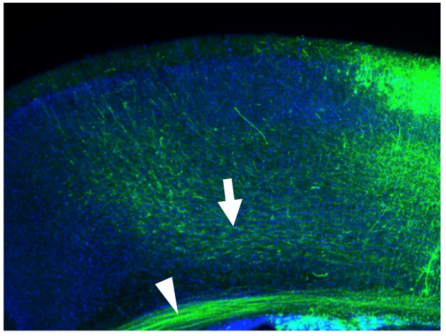
Figure 2. Trace of axonal fiber bundles in the mouse brain.
A cross-sectional image of the mouse cerebral cortex in enlargement. The same method was applied as in the ferret. The cerebral cortex is shown in blue and some neurons are dyed green with GFP. In addition to the deep axonal fiber layer (green, indicated by an arrowhead), a small number of axonal bundles (green, indicated by an arrow) are found. It is considered that in the ferret, these bundles have evolved to be more robust to become the axonal fiber layer on the surface side.
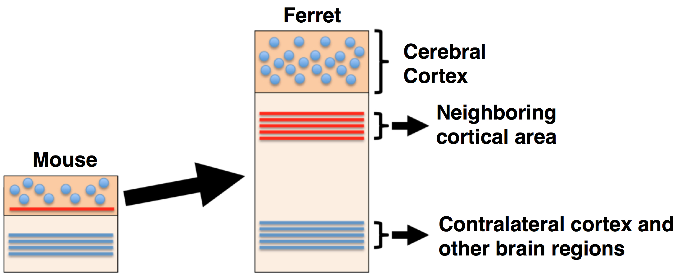
Figure 3. Summary of the present study.
The trace-like axonal fiber bundles are now considered to have become more robust during the evolution of ferrets or humans and, as a result, the second axonal fiber layer has been formed. The destinations of the two axonal fiber layers are also found to be different.
[Glossary]
*1) Cerebral cortex.
The cerebral cortex is the surface region of the cerebrum. The human cerebral cortex is far more developed than that of other animals and is considered to be important in higher brain functions. Damage to the cerebral cortex causes various diseases of the brain as well as psychological disorders. Thus, the cerebral cortex is one of the brain regions receiving extensive attention.
*2) Ferret
The ferret, a higher mammal, is akin to the weasel. The ferret brain is more developed than the mouse brain.
Article
Characterization of the inner and outer fiber layers in the developing cerebral cortex of gyrencephalic ferrets
Journal: Cerebral Cortex
Authors: Kengo SAITO, Keishi MIZUGUCHI, Toshihide HORIIKE, Tung Anh DINH DUONG, Yohei SHINMYO and Hiroshi KAWASAKI
DOI: 10.1093/cercor/bhy312
Funders
Grants-in-Aid for Scientific Research from MEXT, Mitsubishi Foundation, Takeda Science Foundation, and Kanazawa University SAKIGAKE Project 2018.