


Abstract: Tile patterns in which the same shape is laid out without gaps are found in the compound eyes of insects. Hexagonal tile patterns are common while shrimp eyes have a square pattern. We investigated tile pattern formation using Drosophila and revealed that the compound eye tile pattern is controlled by a geometrical division mechanism, Voronoi tessellation, in addition to physical constraints determined by the combination of the regular distribution and growth of the individual eyes.
Tile patterns in which the same shape is laid out without gaps can be seen on castle walls and chess boards in artificial objects, and in insect compound eyes and beehives in biology. Square tiles are common in artificial objects, while hexagonal tiles occur in living things. It was thought that this was due to the physical characteristics of the hexagon being structurally robust, the circumference of each tile being short, and the degree of space filling being high.
It is known, however, that the compound eyes of shrimps and lobsters show a square tile pattern and that those of mantis shrimps consist of a tile pattern in which squares and hexagons are mixed. The compound eye of the fly, Drosophila, also usually shows a hexagonal tile pattern, but in some mutants the pattern changes to a square-type. From these observations, it is considered that the tile pattern of compound eyes is not controlled only by their physical stability. However, it was unknown how such a tile pattern is determined and by what mechanism the hexagonal and square patterns are switched from one to another.
[Results]
This study was performed by a research team led by Prof. Sato of Kanazawa University with the participation of researchers from Hokkaido University, Toyama University and Salesian Polytechnic. The compound eye of wild-type Drosophila individuals shows a hexagonal tile pattern, which in some mutants changes to a square tile pattern (Fig. 1). Because the compound eye is smaller in the square-type mutant, the team suspected that the size of the compound eye itself might affect the tile pattern. Since the compound eye was smaller in the dorsoventral (vertical) direction in this mutant, they thought that the compound eye tissue might be pulled and stretched in the vertical direction by being connected with the head. In fact, it was found that the tension in the vertical direction was increased in the small compound eye of the square-type mutant, and the tissue of the compound eye was elongated in the vertical direction (Fig. 1). However, simply pulling the compound eye in the vertical direction should end up with the hexagons only becoming vertically stretched but could not explain the reason why the hexagons changed to squares.
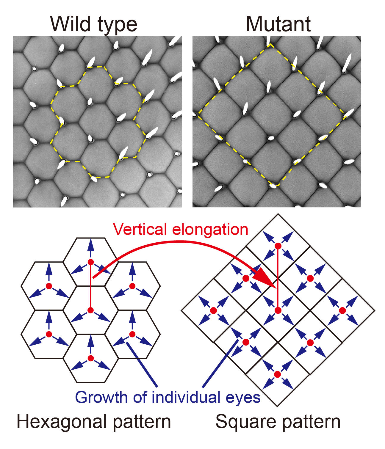
Figure 1. Change from hexagonal to square pattern
A geometrical method called Voronoi tessellation can divide a region evenly around multiple mother points on a plane. This is used, for example, when deciding the appropriate school district for an elementary school (Fig. 2). The perpendicular bisector to the line segment connecting the mother points (elementary schools) constitutes the Voronoi boundary, which divides all areas (school districts) evenly. In the present study, the research team found that not only wild-type hexagonal tiles but also square tiles of mutants are accurately reproduced by Voronoi tessellation. Although it is unreasonable to think that the geometrical method of drawing a perpendicular bisector is performed in living organisms, the same Voronoi boundary is formed when each mother point grows concentrically and collides with another, thus stopping growth (Fig. 2). The team performed experiments and computer simulation to prove that the cells that make up the individual eye produce the same effect as concentric growth by inflating like a balloon.
In this study, the team has shown that the tile pattern is determined by the arrangement of the individual eyes and their concentric growth, that the individual eyes show a hexagonal tile pattern if they are evenly distributed in the vertical and horizontal directions, and that the pattern changes to square-type when the compound eye extends in the vertical direction and the distance between the individual eyes in the vertical direction widens (Fig. 1). It was known that the morphology of cells and tissues is controlled by the function of genes and physical constraints. This study reveals that there is also a geometrical partitioning mechanism, and that these functions are coordinated to control a compound eye tile pattern.
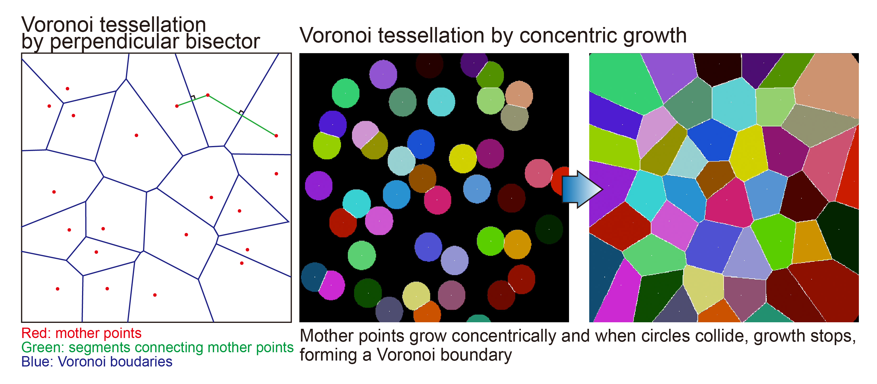
Figure 2. Tile pattern formation by Voronoi tessellation
[Significance of this study]
In developmental biology, synthetic biology, regenerative medicine, etc., it is important to understand the mechanisms that control the morphology of cells and tissues. The results of this study indicate that geometrical patterns based on the growth of concentric circles play an important role in pattern formation in living organisms. Since similar tiling patterns are also found in the columnar structures of the brain, the hepatic lobules of the liver and the auditory epithelium of the inner ear, similar mechanisms may play important roles in a wide variety of tissues as well. Additionally, the optical properties of the visual systems of living organisms are being utilized in new technologies such as artificial compound eyes, for example. It is expected that the results of this study will be applied to bioengineering-related research such as artificial tissues and organs in coming years.
[Funder]
CREST from JST, Grants-in-Aid for Scientific Research from JSPS, MEXT, Takeda Science Foundation, Uehara Memorial Foundation, and Cooperative Research of ‘‘Network Joint Research Center for Materials and Devices’’
[Fund Number]
JPMJCR14D3, 18K06251, 18K13452, 15H05857, 19K03611, 20H05948, 17H03542, 17H05739, 17H05761, and 19H04771.
[Article]
Title: Tiling mechanisms of the Drosophila compound eye through geometrical tessellation
Journal: Current Biology
Authors: Takashi HAYASHI, Takeshi TOMOMIZU, Takamichi SUSHIDA, Masakazu AKIYAM, Shin-Ichiro EI, Makoto SATO
DOI: 10.1016/j.cub.2022.03.046