- Home
- Highlights
- Walking proteins caught on film
Highlights
Walking proteins caught on film
The first high-resolution video footage showing the movement of individual protein molecules has been acquired by Japanese researchers using high-speed atomic force microscopy (HS-AFM).
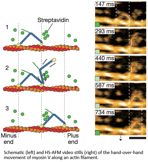
The research team, led by Toshio Ando at Kanazawa University’s Department of Physics and funded by the Japan Science and Technology Agency’s CREST program, produced movies of a two-headed motor protein called myosin V, which transports cargo within cells. Myosin V is believed to move along actin filaments – the internal scaffolding that strengthens cell structures– via a bipedal walker-like hand-over-hand mechanism.
Previous studies using methods such as electron microscopy, x-ray crystallography and nuclear magnetic resonance recording have only produced static images of this dynamic protein behaviour. Fluorescence microscopy has provided moving images, but only of fluorescent particles bound to the proteins, not the proteins themselves. By using HS-AFM, Ando and co-workers were able to simultaneously record the structure and dynamics of myosin V molecules moving along actin filaments.
Initially the steps in myosin movement were too fast for the HS-AFM imaging to keep up with, but by adding streptavidin molecules to act as obstacles, the researchers slowed down the motion to manageable speeds. The movies confirmed existing theories about myosin V movement, while revealing some new and interesting mechanisms. For example, the molecules perform sudden ‘foot stomps’ onto the actin that power a forward swing of their protein arm. The swinging is sometimes preceded by an unwinding of the coiled tail of the molecule, suggesting there is internal tension in the molecule that assists with the power stroke.
This direct visualization could be applied to many other protein systems in order to further our understanding of biomolecular action.
Publication and Affiliation
Noriyuki Kodera1,2, Daisuke Yamamoto1,2, Ryoki Ishikawa3 & Toshio Ando1,2,* Video imaging of walking myosin V by high-speed atomic force microscopy. Nature, 468, 72- 76, (2010).
Link
1. Department of Physics, Kanazawa University, Kakuma-machi, Kanazawa 920-1192, Japan
2. CREST, JST, Sanban-cho, Chiyoda-ku, Tokyo 102-0075, Japan
3. Department of Molecular and Cellular Pharmacology, Gunma University Graduate School of Medicine, 3-39-22 Showa-machi, Maebashi 371-8511, Japan
*corresponding author, e-mail address: tando@staff.kanazawa-u.ac.jp
ID: 201212B001